Human Chest Muscles Anatomy : Paint Draw Paint, Learn to Draw: Anatomy Basics: The chest ... : Some people, specifically most men only targets the chest and workouts it in an extreme condition that might lead to asymmetrical figure to your body.
Human Chest Muscles Anatomy : Paint Draw Paint, Learn to Draw: Anatomy Basics: The chest ... : Some people, specifically most men only targets the chest and workouts it in an extreme condition that might lead to asymmetrical figure to your body.. The pectoralis major muscles (also known as the pecs) are located on the front of the rib cage. Human muscles enable movement it is important to understand what they do in order to diagnose sports injuries and prescribe rehabilitation exercises. Anatomical diagram showing a front view of muscles in the human body. These are usually called pectorals. All these organs and muscles function together to ensure proper body function.
Some people, specifically most men only targets the chest and workouts it in an extreme condition that might lead to asymmetrical figure to your body. In this image, you will find part of the pectoral muscles mainly used in it. 1/2 medial of the anterior border of the clavicle, anterior face of the sternum, external face of the 1st to 6th costal cartilage and aponeurosis of the source: How tropomyosin and troponin regulate muscle contraction. A massive chest anchors the upper body and enhances the.
Muscles of the Thoracic Wall - Chest Muscles Anatomy ... from i.pinimg.com In this article, we shall learn about the anatomy of the muscles of the anterior chest. Click on the labels below to find out more about your muscles. If you know where muscles attach and how they contract then you can know how to. 1/2 medial of the anterior border of the clavicle, anterior face of the sternum, external face of the 1st to 6th costal cartilage and aponeurosis of the source: Tough connective tissue at the bottom of the calf muscle merges with the achilles tendon. Atlas of anatomy of the human body: The chest wall is comprised of skin, fat, muscles, and the thoracic skeleton. It provides protection to vital organs (eg, heart and major vessels, lungs, liver) and provides stability for.
Chest muscles is one of the large part muscles in your body that you also need to work out on aside from your arms, legs and core. This is a table of skeletal muscles of the human anatomy. These are usually called pectorals. Attached to the bones of the skeletal system are about 700 named. The muscular system is responsible for the movement of the human body. Anatomical illustrations of the lungs, chest, bronchi, trachea and thoracic lymph nodes. Anatomy of a human body we study anatomy. Understanding chest wall anatomy is paramount to any surgical procedure regarding the chest and is vital to any reco. The gastrocnemius and soleus muscles taper and merge at the base of the calf muscle. All about the chest muscles. Atlas of anatomy of the human body: 15 photos of the chest muscles anatomy. Abdominal muscles anatomy, back muscles anatomy, chest muscles anatomy for bodybuilders anatomy woman, chest muscles pain, chest workouts, pectoralis major anatomy, shoulder muscles anatomy, human muscles, abdominal muscles.
The muscular system is responsible for the movement of the human body. The chest anatomy includes the pectoralis major, pectoralis minor and the serratus anterior. The pectoralis major muscles (also known as the pecs) are located on the front of the rib cage. 15 photos of the chest muscles anatomy. They are the pectoralis major, pectoralis minor, and the serratus anterior.
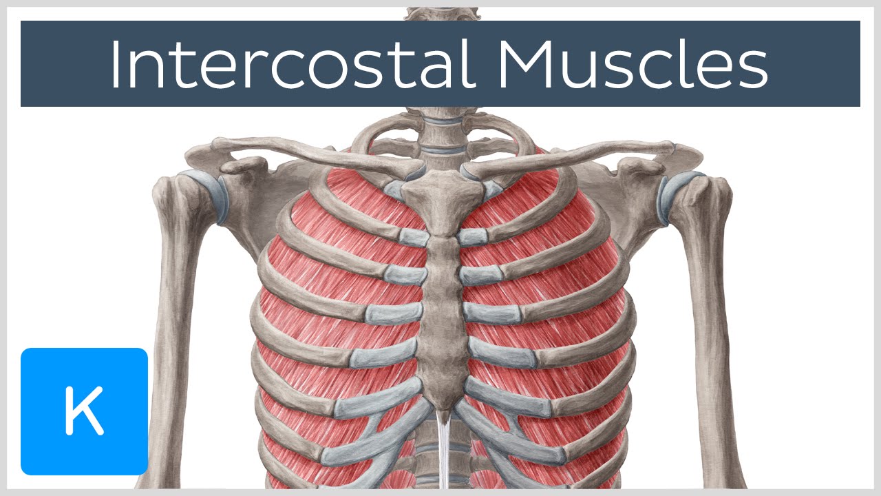
Intercostal Muscles - Function, Area & Course - Human ... from i.ytimg.com This page provides an overview of the chest muscle group. Anatomy of a human body we study anatomy. The chest anatomy includes the pectoralis major, pectoralis minor and the serratus anterior. Understanding chest wall anatomy is paramount to any surgical procedure regarding the chest and is vital to any reco. Chest is a muscle party to which practitioners often attach great importance to the training plan. The gastrocnemius and soleus muscles taper and merge at the base of the calf muscle. Although three ligaments protect and surround the shoulder joint, most of its stability comes from the powerful muscles and tendons of the rotator cuff. Learn about chest muscles human anatomy with free interactive flashcards.
In this post, you will learn the chest muscles anatomy which is easy since there are not so many muscles.
State the action of the major muscles of the human body. These are usually called pectorals. 1/2 medial of the anterior border of the clavicle, anterior face of the sternum, external face of the 1st to 6th costal cartilage and aponeurosis of the source: In this image, you will find part of the pectoral muscles mainly used in it. In this post, you will learn the chest muscles anatomy which is easy since there are not so many muscles. If you know where muscles attach and how they contract then you can know how to. Atlas of anatomy of the human body: 15 photos of the chest muscles anatomy. The chest wall is comprised of skin, fat, muscles, and the thoracic skeleton. Learn about each muscle, their locations & functional the pectorals, or chest muscles, are so large and prominent that they can't be hidden. Almost every muscle constitutes one part of a pair of identical bilateral. Adducts & flexes the arm (humerus). Anatomy of a muscle cell.
The chest anatomy includes the pectoralis major, pectoralis minor and the serratus anterior. Read and learn the following words: Find out more about the individual muscles within the chest anatomy by clicking their respective links throughout this page. You may also find triceps, lateral head brachialis, biceps brachii, latissimus dorsi, deltoid, acromion, clavicle, trapezius, 1st rib, clavicle, acromion, coracoid process, humerus, ulna, radius, sternocleidomastoid. Adducts & flexes the arm (humerus).
Human chest anatomy, illustration - Stock Image - F025 ... from media.sciencephoto.com The muscular system is responsible for the movement of the human body. The shoulder muscles bridge the transitions from the torso into the head/neck area and human muscle system, the muscles of the human body that work the skeletal system, that are under voluntary control, and that are concerned. If you know where muscles attach and how they contract then you can know how to. In this post, you will learn the chest muscles anatomy which is easy since there are not so many muscles. All these organs and muscles function together to ensure proper body function. Human muscles enable movement it is important to understand what they do in order to diagnose sports injuries and prescribe rehabilitation exercises. Attached to the bones of the skeletal system are about 700 named. Read and learn the following words:
All these organs and muscles function together to ensure proper body function.
The gastrocnemius and soleus muscles taper and merge at the base of the calf muscle. These are usually called pectorals. The pectoralis major muscles (also known as the pecs) are located on the front of the rib cage. Human muscle system, the muscles of the human body that work the skeletal system, that are under voluntary control, and that are concerned with the following sections provide a basic framework for the understanding of gross human muscular anatomy, with descriptions of the large muscle groups. Chest muscles is one of the large part muscles in your body that you also need to work out on aside from your arms, legs and core. This page provides an overview of the chest muscle group. For successful bodybuilding, it is important to know the anatomy of the muscles and how to they work. You may also find triceps, lateral head brachialis, biceps brachii, latissimus dorsi, deltoid, acromion, clavicle, trapezius, 1st rib, clavicle, acromion, coracoid process, humerus, ulna, radius, sternocleidomastoid. Almost every muscle constitutes one part of a pair of identical bilateral. How tropomyosin and troponin regulate muscle contraction. Find out more about the individual muscles within the chest anatomy by clicking their respective links throughout this page. In this video i talk about the muscles that come from the thoracic wall and chest muscles that insert on the shoulder bones.✅. Some people, specifically most men only targets the chest and workouts it in an extreme condition that might lead to asymmetrical figure to your body.
In this video i talk about the muscles that come from the thoracic wall and chest muscles that insert on the shoulder bones✅ chest muscles anatomy. This page provides an overview of the chest muscle group.


0 Komentar